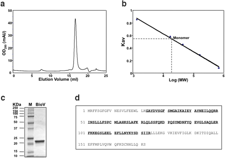
Figure 3. Purification and structural characterization of BioV.
(a) Gel exclusion chromatographic profile of the hexahistidine-tagged BioV analysed on a Superdex 200HR 10/30 column (GE Healthcare) eluted at 0.4 ml min−1. BioV was monitored at 280 nm and eluted at 16.78 min. OD280, optical density at 280 nm; mAu, milli-absorbance units. (b) Determination of BioV solution structure according to elution patterns of a series of standards (Bio-Rad). The standards were vitamin B12 (1.35 kDa), myoglobin (horse, 17 kDa), ovalbumin (chicken, 44 kDa), γ-globulin (bovine, 158 kDa) and thyroglobulin (bovine, 670 kDa). The elution position of BioV gave an estimated molecular mass of 20 kDa based on graphic analysis of the standard curve. Kav, partition coefficient. (c) SDS-PAGE analysis of the purified BioV. The apparent molecular weight of His-tagged BioV is about 21 kDa. M: Molecular weight. (d) Mass spectrometric identification of BioV. The matching peptides are given in bold and underlined type.

