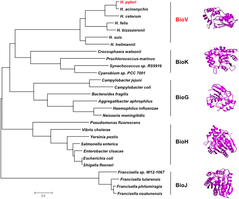
Figure 7. Phylogeny and structural modelling of the bacterial esterases that cleave pimeloyl-ACP methyl ester.
Phylogenetic analyses were conducted by the minimum-evolution method using MEGA6. On the right side of the figure the modelled structure of each esterase is shown together with the known structure of BioH14,15. H. pylori BioV is coloured red.

