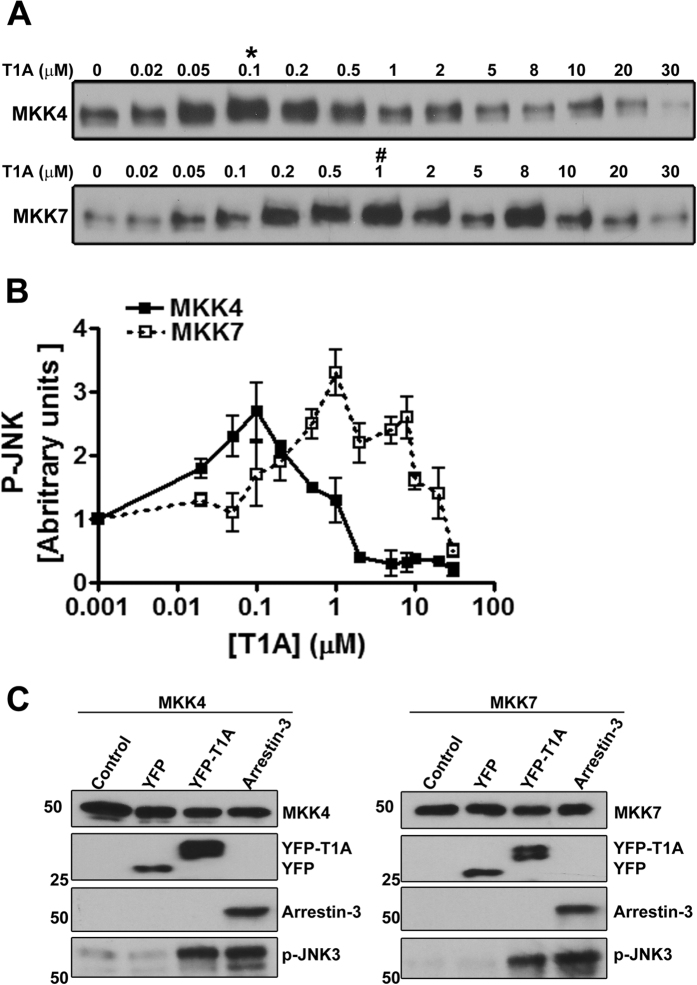
Figure 2. T1A facilitates JNK3 phosphorylation by MKK4 and MKK7.
(A) Representative autoradiograms showing JNK3α2 phosphorylated by purified MKK4 (upper panel) and MKK7(lower panel) at the indicated concentration of synthetic purified T1A peptide (10-s incubation). (B) Quantification of JNK3α2 phosphorylation by MKK4 and MKK7. (C) JNK3α2 phosphorylation by MKK4 and MKK7 in COS7 cells co-expressing JNK3α2 with MKK4 or MKK7 (control) and YFP, YFP-T1A, or arrestin-3. Full blots are shown in Supplemental Fig. S2.

