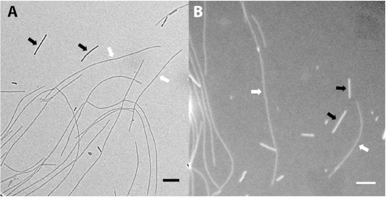
Figure 3. Bright field and dark field imaging of unstained fibrils.
Images of wild type α-synuclein and TMV standards made with (A) bright field and (B) dark field illumination. Examples of TMV standards and amyloid fibrils are indicated by black and white arrows respectively. Scale bars are 200 nm.

