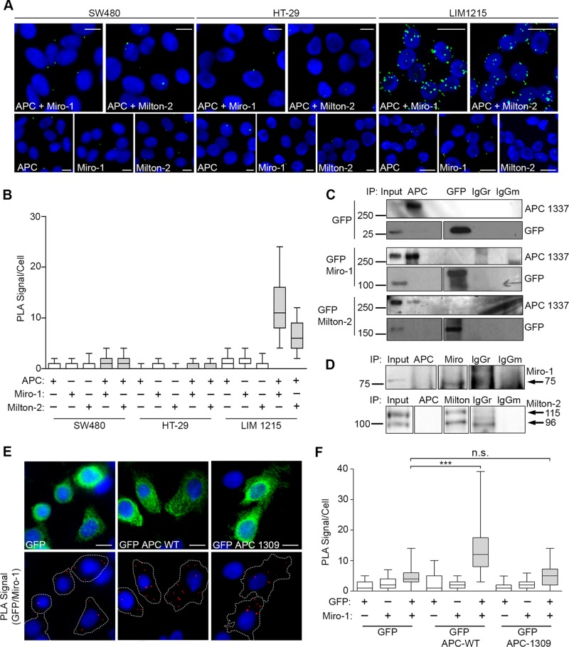
FIGURE 7:
Truncated APC does not interact with the Miro/Milton complex. (A and B) Test for interaction between APC and Miro/Milton in colon cancer cell lines SW480 (APC1377), HT-29 (APC853/1555), and LIM1215 (APC WT) in situ using Duolink PLA. (A) Representative cell images (green dots are positive signals) were quantified in the graph (B). The interaction between APC and Miro/Milton is lost when APC is truncated (SW480 and HT-29 CRC cells) but remains intact when APC is full length (LIM1215 CRC cells). (C) SW480 cells expressing GFP, GFP-Miro-1, or GFP-Milton-2 were subjected to IP assays using anti-APC to pull down the respective GFP-tagged protein and anti-GFP to pull down APC. No bands were detected. Blots were spliced where indicated. (D) SW480 cell lysates were used for IP of endogenous protein; however, antibodies against APC were unable to pull down endogenous Miro-1 or Milton-2. Blots were spliced where indicated. (E) SW480 cells transiently expressing GFP-APC-WT and GFP-APC-1309 were subject to Duolink PLA where interactions between GFP-tagged APC sequences and Miro-1 were observed in situ by immunofluorescence microscopy. (F) Quantification of PLA signals revealed a significant positive signal above GFP background for GFP-APC-WT that was not observed for the GFP-APC mutant (***, p < 0.001; n.s., not significant). n > 125 per sample over two independent experiments. Box-and-whisker plot presented as a median (line), upper/lower quartile (box), and min/max (error bars with 95% CI). Statistical analysis by Mann-Whitney U-test with Bonferroni correction. Scale bars: 10 μm.

