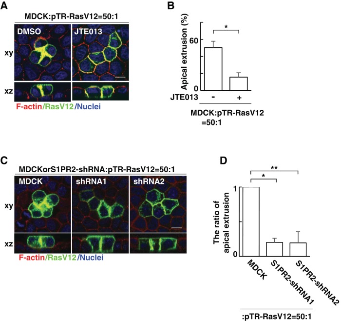
FIGURE 1:
S1PR2 in the surrounding normal cells plays a positive role in the apical extrusion of RasV12-transformed cells. (A) Confocal microscopic immunofluorescence images of xy- and xz-sections of MDCK-pTR GFP-RasV12 cells in a monolayer of normal MDCK cells cultured in the absence or presence of JTE013. Twenty-four hours after tetracycline addition, cells were stained with phalloidin (red) and Hoechst (blue). (B) Quantification of the apical extrusion of RasV12 cells. Data are mean ± SD from three independent experiments. *p = 0.0027. (C) Confocal microscopic immunofluorescence images of xy- and xz-sections of MDCK-pTR GFP-RasV12 cells in a monolayer of normal MDCK cells or S1PR2-knockdown MDCK cells. Cells were stained with phalloidin (red) and Hoechst (blue). Scale bars, 10 μm (A, C). (D) Quantification of the apical extrusion of RasV12 cells. Data are mean ± SD from three independent experiments. Values are expressed as a ratio relative to MDCK cells. *p = 2.2 × 10⁻⁵, **p = 0.0010.

