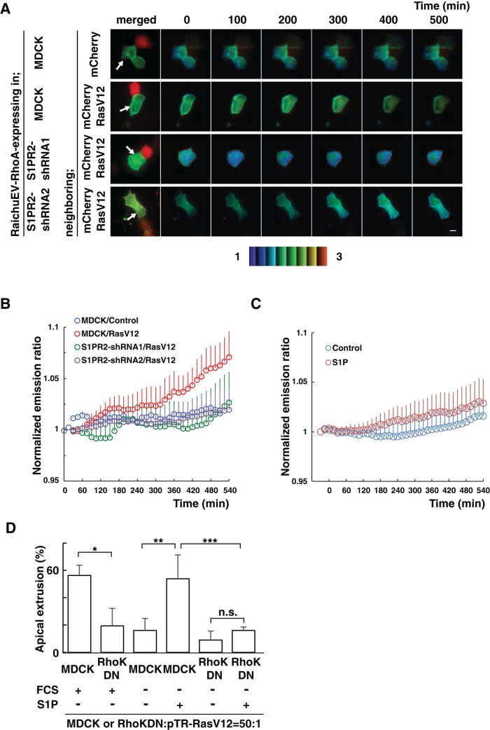
FIGURE 4:
The S1P–S1PR2 pathway acts upstream of Rho–Rho kinase in normal cells neighboring RasV12-transformed cells. (A) MDCK cells or S1PR2-knockdown MDCK cells were transfected with an expression vector for the FRET-based biosensor RaichuEV-RhoA. The cells were further transfected with an expression vector for mCherry alone or together with that for RasV12 and then seeded on the collagen gel. After 12 h, the sample was screened under a fluorescence microscope for a pair of adjacent cells, one of which expressed RaichuEV-RhoA and the other mCherry/RasV12. The Raichu-expressing cells were then monitored by dual-emission fluorescence microscopy. FRET/CFP ratio images were generated to represent FRET efficiency. Representative images at the indicated time points. The upper and lower limits of the ratio range are indicated at the bottom. Arrows indicate Raichu-expressing cells neighboring mCherry-expressing cells. Scale bar, 10 μm. (B, C) Traces for three-point moving average of the FRET/CFP emission ratio. (B) Quantification of A. p = 5.2 × 10⁻⁵ between MDCK/control and MDCK/RasV12; p = 4.0 × 10⁻⁸ between MDCK/RasV12 and S1PR2-shRNA1/RasV12; p = 2.0 × 10⁻⁶ between MDCK/RasV12 and S1PR2-shRNA2/RasV12. n = 7, MDCK/control; n = 5, MDCK/RasV12; n = 8, S1PR2-shRNA1/RasV12; and n = 8, S1PR2-shRNA2/RasV12. (C) Effect of S1P on the Rho activity in normal cells. p = 1.7 × 10⁻⁹ between MDCK/control and MDCK/RasV12. n = 6, control (fatty acid–free BSA); and n = 11, 200 nM S1P. Error bars indicate SEM. Time 0 min is set when the microscopic observations start (A, B) or S1P is added (C). (D) Effect of exogenously added S1P on the apical extrusion of RasV12 cells surrounded by normal MDCK cells or MDCK cells expressing the Rho kinase dominant-negative mutant. Data are mean ± SD from three (first, second, fifth, and sixth bars) or six (third and fourth bars) independent experiments. *p = 0.013, **p = 0.00082, ***p = 0.0088. n.s., not significant.

