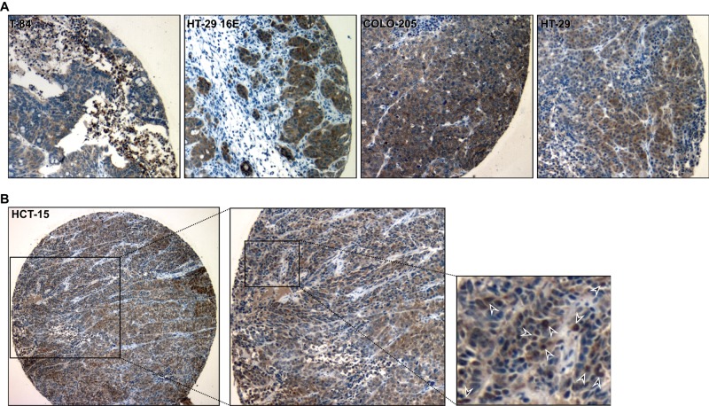
FIGURE 4:
Nuclear localization of villin in colon cancer cell xenografts. Colon cancer cell lines endogenously expressing villin—T-84, HT-29 16E, COLO-205, HT-29 19A, and HCT-15 cells—were grown as xenografts in SCID mice. The tumors were excised and Formalin fixed. Villin expression was determined by immunohistochemistry using a commercially available anti-villin antibody. (A) Representative images of T-84, HT-29 16E, COLO-205, and HT-29 19A. (B) Higher magnification of HCT-15 xenograft shows localization of villin in individual nuclei (arrowheads).

