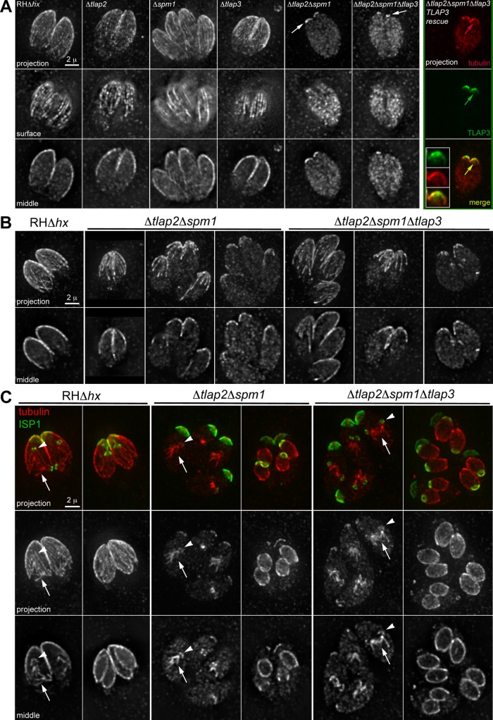
FIGURE 10:
The coating proteins protect the stability of cortical microtubules as an ensemble. (A) Deconvolved wide-field images of cold-treated parental RHΔhx parasites and various coating protein–knockout mutants labeled with a rabbit anti–Tgβ-tubulin antibody (Morrissette and Sibley, 2002b). Intracellular parasites were incubated at 8°C for 3.5 h before processing for immunofluorescence. In the Δtlap2Δspm1Δtlap3-TLAP3 rescue parasites, mNeonGreenFP-TLAP3 expression was driven by the 2-kb genomic region immediately upstream of tlap3 in the plasmid pTKO2_II_mNeonGreenFP-TLAP3. Insets, 2×. Arrows indicate residual cortical labeling by the anti–Tgβ-tubulin antibody in the Δtlap2Δspm1, Δtlap2Δspm1Δtlap3, or Δtlap2Δspm1Δtlap3- TLAP3 rescue parasites. (B, C) Deconvolved wide-field images of intracellular parental RHΔhx, Δtlap2Δspm1, and Δtlap2Δspm1Δtlap3 parasites labeled with a rabbit anti–Tgβ-tubulin antibody. The parasites were cultured under standard culture conditions (i.e., with no cold treatment) before processing for immunofluorescence. (B) Interphase parasites. (C) Parasites with forming daughters (arrows, mitotic spindle; green, anti-ISP1; red, anti–Tgβ-tubulin).

