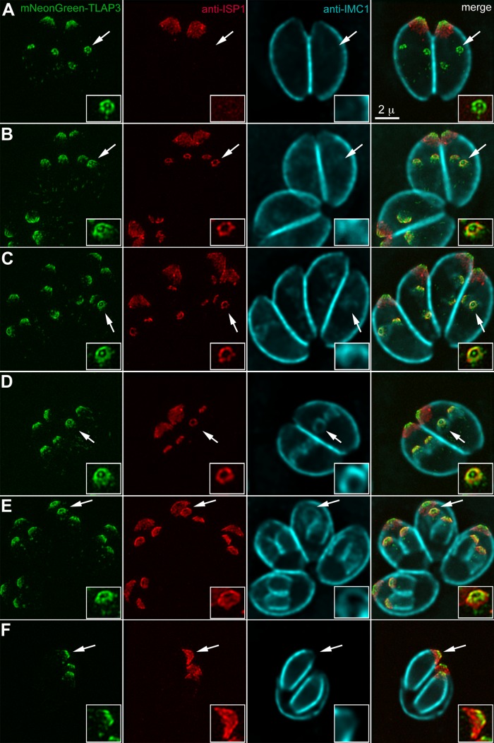
FIGURE 3:
TLAP3 decoration of the cortical microtubules is established early during daughter construction. (A–F) mNeonGreenFP-TLAP3 knock-in parasites at different stages of the cell cycle labeled with a mouse anti-ISP1 antibody and a rabbit anti-IMC1 antibody. The mNeonGreenFP-TLAP3 and anti-ISP1 images are projections of 3D-SIM images. The anti-IMC1 images, visualized by a goat anti-rabbit Cy5 antibody, are projections of deconvolved wide-field images, as the Cy5 fluorophore is not compatible with SIM imaging. Surface sections were not included in the anti-IMC1 projections to better display the signal from daughter cells. The Cy5 channel was misregistered slightly with the other two channels. Alignment was done by manually lining up the edge of ISP1 and IMC1 labeling. Insets (2×) are contrast enhanced and include regions of daughter parasites indicated by the arrows. For clarity, the IMC1 channel is not shown in the insets of the merged image panels. Green, mNeonGreenFP-TLAP3. Red, anti-ISP1. Cyan, anti-IMC1. Scale bars, 2 μm.

