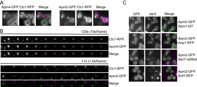
FIGURE 2:
Apm2 localizes to late Golgi/early endosomes. (A) Fluorescence microscopy of live endocytosis-defective sla2 yeast shows that Apm2 is not present at cell-surface clathrin-coated pits. White arrows indicate puncta that colocalize in GFP and RFP channels. (B) Kymograph analysis of Apm4-GFP or Apm2-GFP relative to cortical clathrin patches marked by Clc1-RFP. Cells were incubated with the actin inhibitor latrunculin A and imaged by TIRF microscopy. (C) Localization of Apm2-GFP relative to that of Apm1-tdTomato (AP-1 complex), Anp1-RFP (early Golgi), Sec7-DsRed (late Golgi/endosomes), and Snf7-RFP (late endosomes). Scale bar, 4 μm.

