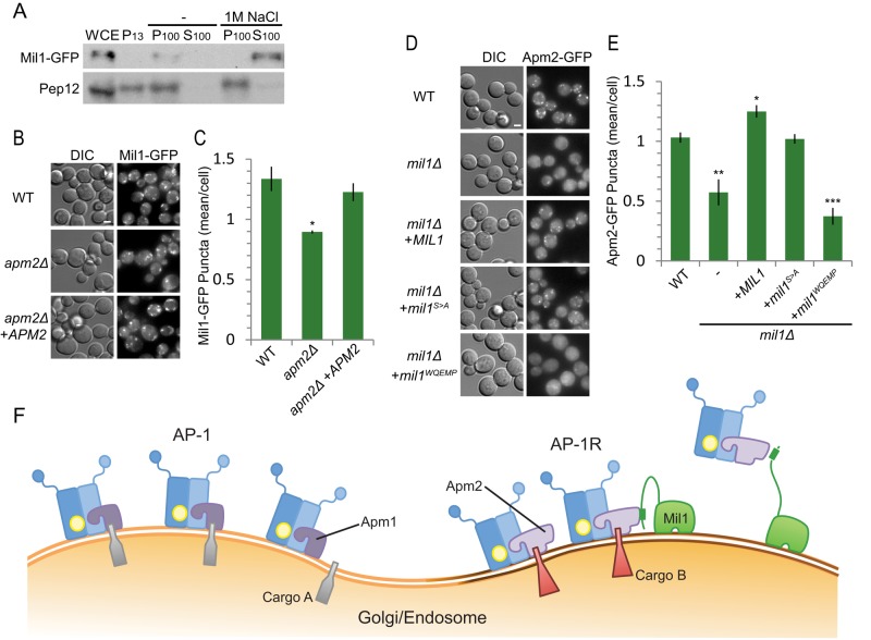
FIGURE 6:
Mil1 is a peripheral membrane protein that promotes Apm2 membrane recruitment. (A) Subcellular fractionation of Mil1 and the transmembrane protein Pep12. WCE, whole-cell extract. P13, low-speed pellet fraction; P100, high-speed pellet fraction; S100, high-speed soluble fraction. Samples were resolved by 15% SDS–PAGE and detected by immunoblotting. (B) Representative maximum-intensity z-projection images from nine slices at 0.3-μm increments. Scale bar, 2 μm. (C) Localization of Mil1-GFP in wild-type or apm2 mutant strains quantified in a single slice. Unpaired t test, *p < 0.05. Error bars represent SEM (n = 3). (D) Representative maximum-intensity z-projection images from nine slices at 0.3-μm increments. Scale bar, 2 μm. (E) Localization of Apm2-GFP expressed in wild-type or mil1 mutant strains and quantified in a single slice. Unpaired t test, ***p < 0.0001, **p < 0.01, and *p < 0.05. Error bars represent SEM (n = 4). (F) Proposed model: Mil1 helps to recruit Apm2 to a distinct membrane region.

