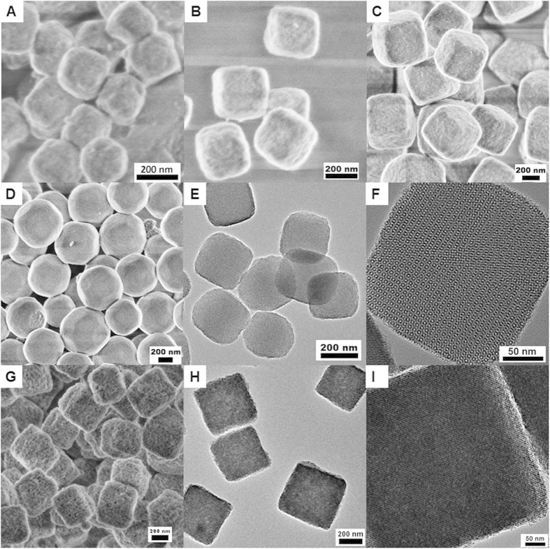
Figure 2. The characterization of the PMO nanocubes with electron microscopy.
FESEM images (A–D) of the PMO nanocubes and truncated-cubes with sizes of 150 nm (A), 250 nm (B), 400 nm (C) and 600 nm (D); TEM images for the PMO nanocubes with a size of 250 nm (E,F); HRSEM (G) and TEM (H,I) images of the PMO nanocubes with a size of 250 nm after hydrothermal treatment at 120 °C.

