FIG 6.
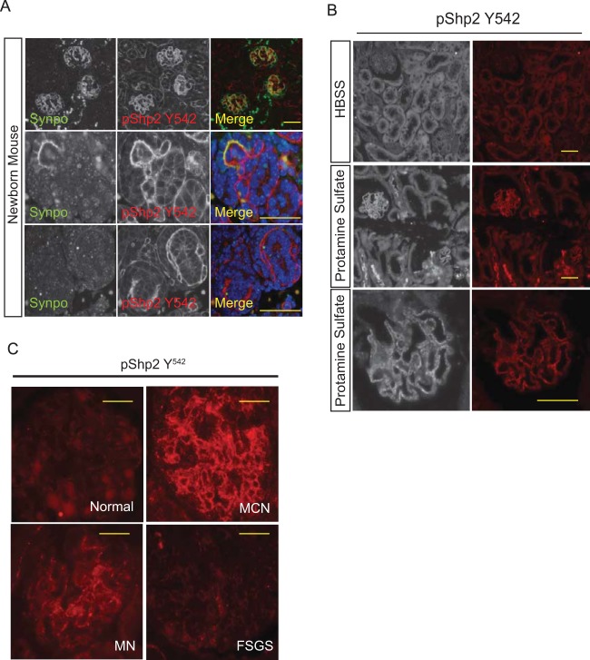
(A) Shp2 is tyrosine phosphorylated in developing glomeruli. Newborn mouse kidney sections were prepared as described in Materials and Methods. The sections were analyzed by indirect immunofluorescence microscopy following immunostaining with p-Shp2 (Y542) antibody. Synaptopodin antibody was used to identify podocytes in the section. (Top row) Lower-magnification images of kidney cortex. (Bottom rows) Higher-magnification images of developing glomeruli. Scale bars, 20 μm. (B) Shp2 is tyrosine phosphorylated following perfusion with protamine sulfate. Adult mouse kidneys were perfused with HBSS and protamine sulfate prior to perfusion fixation with paraformaldehyde. Kidney sections were analyzed by indirect fluorescence microscopy. Magnification, ×200 (top 2 rows) and ×400 (bottom row). Scale bars, 20 μm. (C) Enhanced Shp2 phosphorylation in human glomerular diseases. Representative images show p-Shp2 Y542 antibody staining of paraffin-embedded kidney sections from healthy subjects and from patients with minimal-change nephrosis (MCN), membranous nephropathy (MN), and focal segmental glomerulosclerosis (FSGS). Kidney biopsy sections from 5 or more patients in each disease category were immunostained. Scale bars, 20 μm.

