
Figure 2.
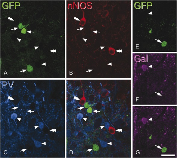
Lack of overlap between green fluorescent protein (GFP) and other markers for inhibitory interneuron populations. Confocal scans of a transverse section from a neuropeptide Y–GFP mouse that had been immunostained for neuronal nitric oxide synthase (nNOS) (red), parvalbumin (PV, blue), and galanin (Gal, magenta). A–D show 3 GFP+ cells (green, arrows) in lamina IIi-III that are not immunoreactive for either nNOS or PV but are surrounded by cells that are either PV-immunoreactive (arrowheads) or nNOS-immunoreactive (double arrowheads). (E–G) Part of lamina II from the same section scanned to reveal galanin and GFP. A GFP+ cell that lacks galanin (arrow) and a galanin-immunoreactive cell that lacks GFP (arrowhead) are shown. A–D consist of 3 optical sections at 1 μm z-spacing, while E–G are from a single optical section. Scale bar = 20 μm.