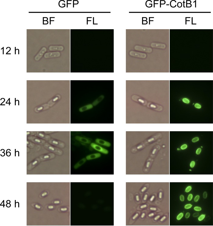
FIG 5.
Bright-field and fluorescence microscopic analysis of GFP-CotB1 during sporulation. The B. cereus wild-type strains carrying pHT-gfp-cotB1 (GFP-CotB1) or pHT-gfp (GFP) were grown in mR2A medium supplemented with 100 μg ml−1 silicate. Samples were taken at the indicated times after inoculation and observed by bright-field (BF) and fluorescence (FL) microscopy.

