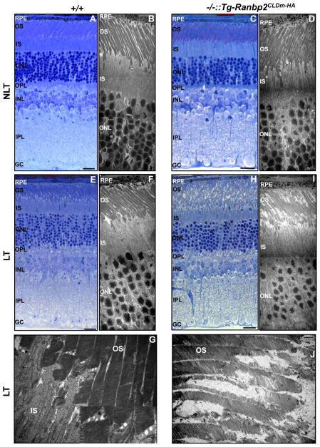
Fig. 2.
Morphological changes of photoreceptor neurons between wild-type and Ranbp2−/−::Tg-Ranbp2CLDm-HA mice reared in the absence and presence of light-stress. There are no visible morphological changes under light (A, C) and ultrastructural microscopy (B, D) of photoreceptors of wild-type (A, B) and Ranbp2−/−::Tg-Ranbp2CLDm-HA mice (C, D) reared under non-light treatment (NLT) at 24 weeks of age (A–D). Under light-treatment (E–J), the OS of photoreceptors of 24-week-old wild-type mice show mild disruption primarily at the base of the OS as shown by light (E) and ultrastructure microscopy (F, G), whereas light (H) and ultrastructure microscopy (I, J) show profuse derangement and erosion of the OS of photoreceptors, respectively, of age-matched Ranbp2−/−::Tg-Ranbp2CLDm-HA mice. Inset picture is magnified dashed box in (G). Legend: OS, outer segments of photoreceptors; IS, inner segments of photoreceptors; ONL, outer nuclear (cell bodies) layer of photoreceptors; OPL, outer plexiform layer; INL, inner nuclear (cell bodies) layer of retinal neurons; IPL, inner plexiform layer; GC, ganglionic neurons; RPE, retinal pigment epithelium; NLT, non-light treatment; LT, light treatment (light-stress); +/+, wild type; −/−::Tg-Ranbp2CLDm-HA, Ranbp2−/−::Tg-Ranbp2CLDm-HA. Scale bars: 20 μm (A, C, E, H), 5 μm (B, D, F, I), 1 μm (G, J).

