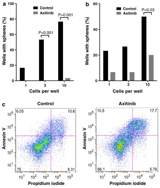
Fig. 2.
Effect of axitinib on GSC clonogenicity and apoptosis in vitro. a, b Secondary sphere formation assay with MGG4 (a) and MGG8 (b) GSCs. Axitinib dose was 1 μM (a) and 0.01 μM (b). Fraction of wells containing sphere(s) is shown for different cell densities (1, 3 and 10 cells per well). c MGG4 GSCs were treated with axitinib (3 μM) or control for 48 hours, stained for Annexin V and propidium iodide, and analyzed by flow cytometry. Numbers in each quadrant denote the percentage of each fraction

