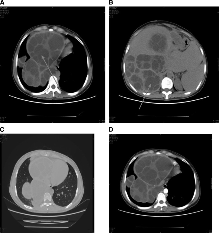
Figure 2.
Plain and enhanced computed tomography (CT) scans from patient no. 2. The CT scans were obtained before the first surgery (A and B) and the second surgery (C and D). (A and B) Plain CT showing multiple hydatids in the abdominal and pelvic cavities and vesicae that were formed as a result of Echinococcus granulosus infection (arrows). (C) Plain and (D) enhanced CTs of the chest.

