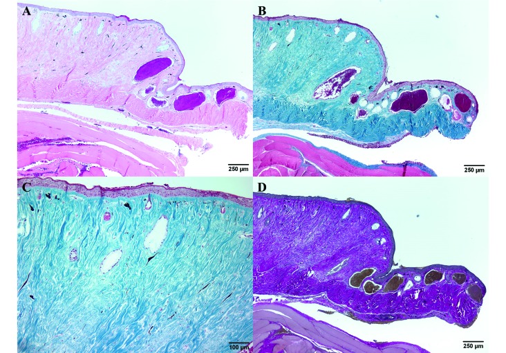
Figure 4.
(A) The dermis is greatly expanded by connective tissue and shows a marked decrease in the number of glands. Hematoxylin and eosin stain. (B) The excessive connective tissue stains blue, indicating that it is composed of collagen. Masson trichrome stain. (C) Higher magnification of panel B. The connective tissue of the dermis expanded by abundant blue-staining collagen; few normal dermal glands remain. Masson trichrome stain. (D) The bright-red staining of the connective tissue with an absence of black-staining material demonstrates the presence of collagen and an absence of elastic fibers. Verhoeff–van Gieson stain.

