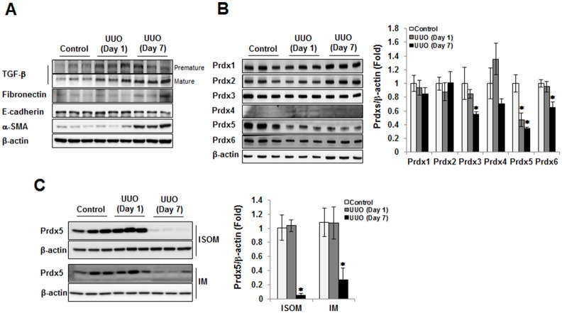
Fig 1. The protein levels of Prdxs in UUO kidney.
To check of progression level of renal fibrosis in left kidney during ureter obstruction, we analyzed the expression of fibrotic marker proteins (premature and mature form of TGF-β, fibronectin, and α-SMA) and epithelial marker protein (E-cadherin) (A). To assess the involvement of Prdxs in renal fibrosis, we analyzed the expression of several Prdx isotypes (Prdx1, Prdx2, Prdx3, Prdx4, Prdx5, and Prdx6) in the cortex/outer stripe of the outer medulla tissue homogenate UUO kidney. Bar graphs show mean Prdxs/β-actin expression as measured by densitometry (B). To further understand the involvement of Prdx5 in renal fibrosis, we analyzed the expression level of Prdx5 in the inner stripe of the outer medulla (ISOM) and inner medulla (IM). Bar graphs show mean Prdx5/β-actin expression as measured by densitometry (C). *p < 0.05 Day 1 and Day 7 vs. control kidney

