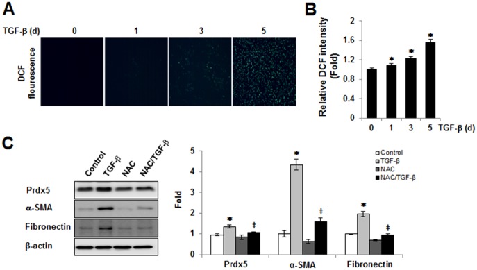
Fig 3. The role of the intracellular ROS in TGF-β induced Prdx5 up-regulation.
To assess TGF-β mediated intracellular ROS levels, ROS probe (10 μM CM-H2DCFDA, Invitrogen) were loaded in TGF-β treated NRK49F cells for 0, 1, 3, and 5 days. Intracellular ROS levels were analyzed using fluorescence microscope (Nikon ECLIPSE TE2000) (A). Bar graphs show relative ROS levels as measured by fluorescence microplate reader (Gemini XPS Microplate Reader) (B). To assess the role of ROS in TGF-β induced Prdx5 up-regulation, NRK49F cells were incubated for 3 day with TGF-β in the presence or absence of 10 mM NAC. The protein levels of Prdx5, fibronectin, and α-SMA were assayed using western blotting (C). *p<0.05 TGF- β treated 1, 3, and 5 day vs. control 0 day

