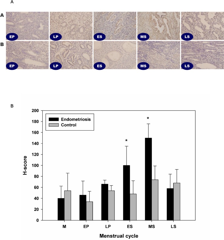
Fig 1. Immunohistochemistry analysis of HMGB-1 expression in the human endometrium.
In control endometrium, HMGB-1 was expressed in the glandular cells of the epithelium and in stromal cells (N = 27). No significant differences were observed according to the menstrual cycle (A). In EM from a patient with endometriosis, significantly increased HMGB-1 expression was detected in the early and mid-secretory phase as compared to the control EM (N = 33, B). There were no significant differences in HMGB-1 expression between the two groups during the proliferative phase. *, P < 0.05 versus the control.

