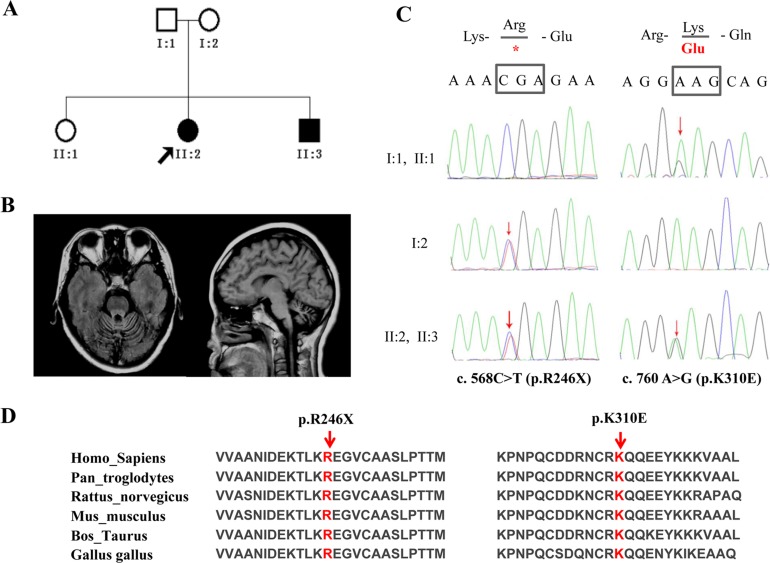
Fig 1. Genetic findings in a family with UBA5 mutations.
(A) The pedigree of family 1 with autosomal recessive spinocerebellar ataxia. (B) Brain magnetic resonance imaging of patient II:2. Panel (left): axial T1-weighted image showing atrophy of the cerebellar vermis. Panel (right): midline sagittal T1-weighted image showing cerebellar atrophy, particularly in the superior vermis, with enlargement of the fourth ventricle. (C) The UBA5 variants [c. 568C > T; c. 760A > G], [p. Arg246X (R246X); p. Lys310Glu (K310E)] segregated in this family. Red arrows indicate the mutation sites. (D) Two mutations (red) affected amino acids that are highly conserved across species.

