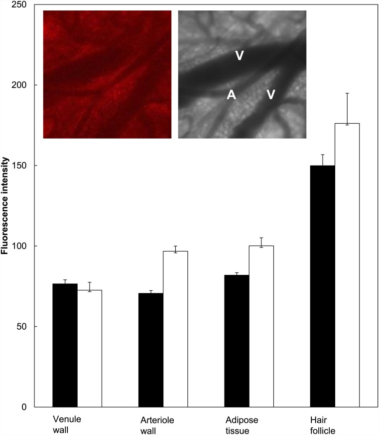
Fig 4. PpIX fluorescence determined using the intra-vitally skin-fold chamber model.
Fluorescence intensity in different locations low in the dermis determined 4 hours after ALA (black) or BF-200 ALA (white) application using intra-vital confocal microscopy and the skin-fold chamber model. Insert shows an example of intra-vital confocal fluorescence microscopy and transmission images (V = venule, A = arteriole).

