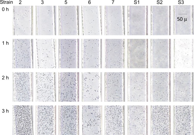
Fig 3. Growth of eight strains of Pseudomonas aeruginosa in the DSTM device over time.
Suspensions of McFarland 0.4–0.5 were used. Strain 2 grew too rapidly to analyze images at 3 h using the software, although it was possible to judge this visually. The images of strains 2, 3, 5, and 7 at 2 h were useful for image analysis, and all images at 3 h except for strain 2 were analyzable using the software.

