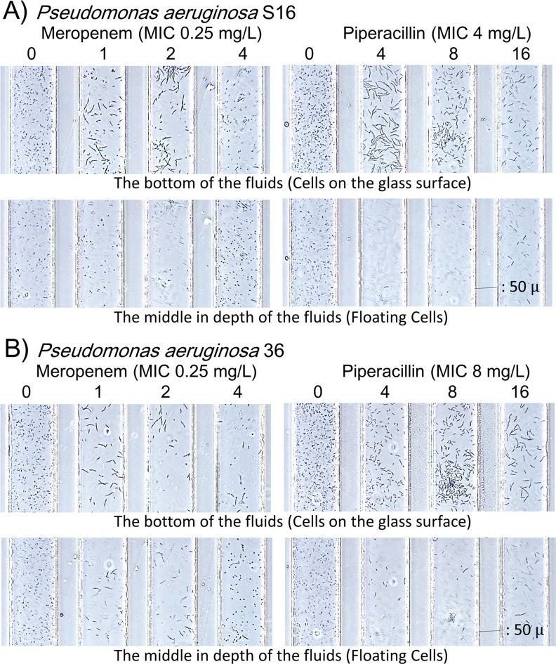
Fig 8. Different image outputs between the bottom and the middle depths of the channels.
Images from A) strain S16 and B) strain 36 at 3 h. Elongated cells were usually located on the glass surface, while spheroplast cells were floating in the fluids. The images focused on the glass surface are preferable for analysis using the software.

