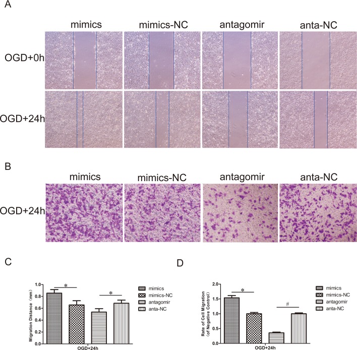
Fig 3. Effect of miR-21 on HUVEC migration.
(A) Scratch wound migration assay in vitro. After transfection with miR-21 mimics, miR-21 mimic negative control, antagomir-21 or antagomir-21 negative control, confluent HUVECs monolayers were exposed to OGD and then scratched to induce horizontal migration of HUVECs (magnification, x100). (C) Comparison of the distance of HUVECs horizontal migration at 0 h and 24 h between treatment groups and negative control groups. Values represent means±S.D; n = 3; *p<0.05. (B) Transwell chamber migration assay in vitro. HUVECs subjected to transfection and OGD mentioned above were trypsinized and seeded into the upper chamber and then allowed to vertically migrate for 24 h. Cells on the lower surface of the membrane were stained with 0.1% crystal violet (lavender) (magnification, x100). (D) Comparison of the rate of cell migration in the treatment groups relative to their respective negative control groups at 24 h. Values represent means±S.D; n = 3; *p<0.05, #p<0.05.

