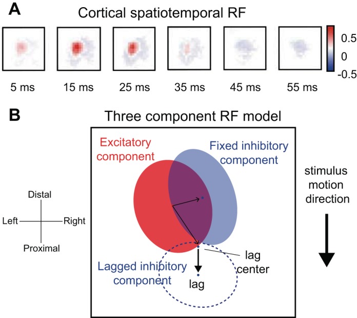
Fig. 5.

Spatial temporal receptive fields (STRFs). A: STRF estimated for a neuron in area 1 using a modified form of linear regression similar to reverse correlation. This STRF comprises a central excitatory region, fixed inhibition, and lagged inhibition on a single finger. Color bar indicates pixel intensities in spikes per second per micrometer. B: 3-component model that includes fixed excitatory (red), fixed inhibitory (solid blue), and lagged inhibitory components (dashed blue). The sum of the elliptical regions yields the full STRF. The centers of the fixed and lagged inhibitory components are indicated by the blue dots. For RFs estimated from responses to moving dot stimuli (see DiCarlo and Johnson, 2000), the location of the lagged inhibitory component, with respect to the excitatory region, depends on the direction in which the stimulus is scanned (thick black arrow). The location of the fixed inhibitory component remains unchanged over motion directions (adapted from Sripati et al. 2006b).
