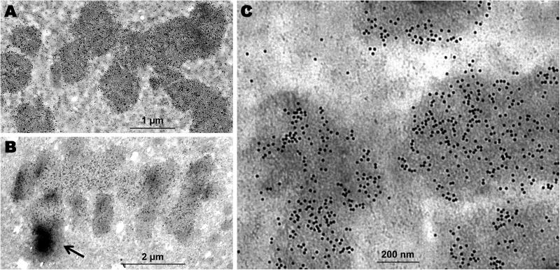
Figure 3.
MCF-7 cells immunogold stained with H3S10ph antibody. (A) The 10 nm immunogold beads distributed relatively evenly along the condensed chromosomes at metaphase. (B) The immunogold bead density distribution displayed dramatic gradients (arrow) among chromatids at anaphase. (C) Immunoelectron microscopy image to show the portion of chromosomes at metaphase with distinctive 10 nm gold beads. A, Scale bar = 1 μm; B, Scale bar = 2 μm; C, Scale bar = 200 nm.

