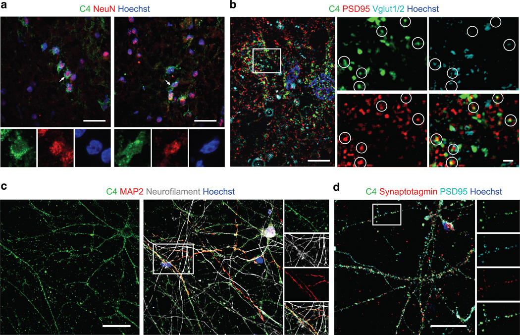
Figure 6. C4 protein at neuronal cell bodies, processes and synapses.
(a) C4 protein localization in human brain tissue. Two representative confocal images (drawn from immunohistochemistry performed on samples from five individuals with schizophrenia and two unaffected individuals) within the hippocampal formation demonstrate localization of C4 in a subset of NeuN+ neurons.
(b) High-resolution structured illumination microscopy (SIM) imaging of tissue in the hippocampal formation reveals colocalization of C4 with the presynaptic terminal marker Vglut1/2 and the postsynaptic parker PSD95.
(c) Confocal images of primary human cortical neurons show colocalization of C4, MAP2, and neurofilament along neuronal processes.
(d) Confocal image of primary cortical neurons stained for C4, presynaptic marker synaptotagmin, and postsynaptic marker PSD95.
Scale bar for a, c, and d = 25 µm; b = 5 µm; b (inset)= 1 µm. Extended Data Fig. 9 contains additional data on antibody specificity

