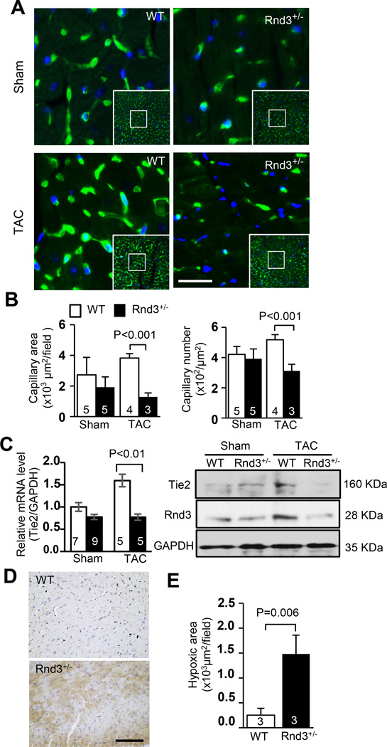
Figure 2. Rnd3 deficiency resulted in an angiogenesis defect in the mouse heart in response to TAC.
(a) Fewer capillaries were observed in the post-TAC Rnd3+/− heart by isolectin staining shown in green. Blue indicated nuclear DAPI staining. Scale bar represents 12.5 µm. (b) Cardiac capillaries were quantified by LAS V4.0 software (Germany). Smaller capillary areas and fewer capillary numbers were observed in the Rnd3+/− mouse heart compared to the WT control after TAC. (c) The responsive increases in angiogenic marker Tie2 mRNA and protein levels were attenuated in the post-TAC Rnd3+/− heart. (d) Larger hypoxic areas were detected in the Rnd3+/− heart compared to the WT heart after TAC. Hypoxyprobe™-1 staining (brown) showed hypoxic myocardium. Scale bar represents 50 µm. (e) Quantification of hypoxic areas by LAS 4.0 software (Germany). The numbers in the columns represented the number of mice in each group.

