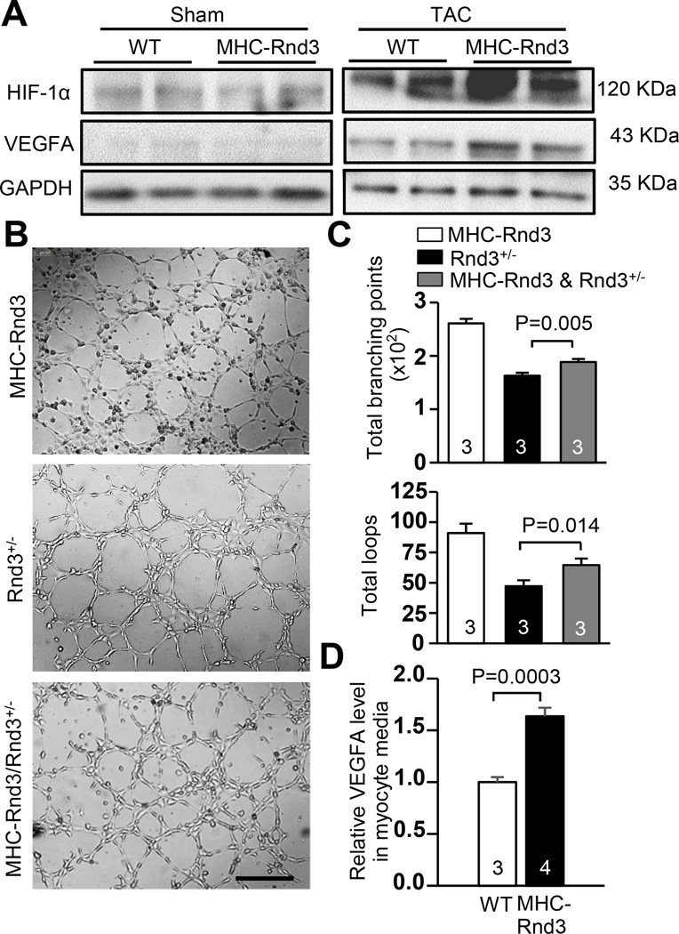
Figure 7. HIF1α-VEGFA signaling was enhanced in MHC-Rnd3 cardiomyocytes.
(a) Upregulation of HIF1α and VEGFA protein levels in the MHC-Rnd3 mouse heart in response to TAC was shown by Western blot analysis. (b) HUVEC cell tube formation was highly promoted by the media from the MHC-Rnd3 cardiomyocytes. The resulting tube formation deficiency from culturing with the Rnd3+/− cardiomyocyte media was partially rescued when co-cultured with the MHC-Rnd3 cardiomyocyte media. Scale bar represents 500 µm. (c) The tube formation was quantified by WimTube software. (d) Cardiomyocytes from the MHC-Rnd3 heart released more VEGFA compared to the control WT cardiomyocytes, indicating strong VEGFA paracrine activity. The numbers in the columns represented the number of mice in each group.

