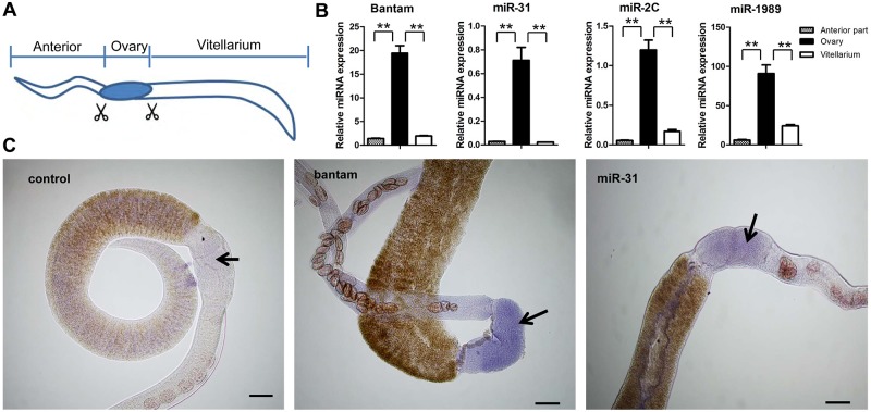
Fig 5. Female enriched S. japonicum miRNAs are predominantly expressed in the ovary.
(A) Schematic diagram of the dissection of female schistosomes. (B) qRT-PCR analyses for the expression of four female enriched miRNAs (miR-31, bantam, miR-1989, miR-2c). * means P ≤ 0.05 and ** means P ≤ 0.01 (student’s t test, ovary vs. anterior and ovary vs. vitellarium). Data illustrate representative findings and show the mean and standard errors derived from triplicate experiments. (C) In situ hybridization experiments were carried out using a labeled locked nucleic acid (LNA) complementary to miR-31 or bantam miRNA. A labeled scrambled LNA was used as a control. Bars indicate 100 μm.

