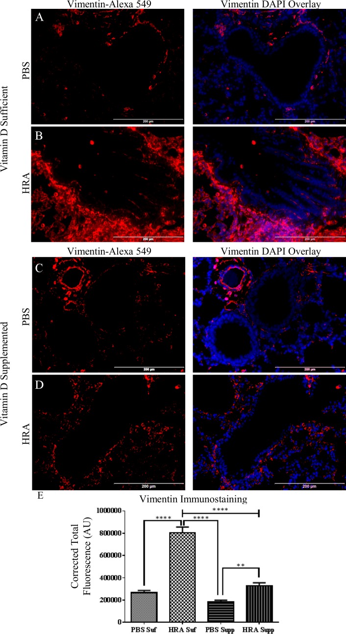
Fig 7. Effect of vitamin D on vimentin expression in lung epithelium of PBS and HRA-sensitized and challenged mice.
(A, B) Immunofluorescence image shows the immunostaining of vimentin, in the lungs of vitamin D-sufficient PBS control mice compared to vitamin D-sufficient HRA-sensitized and challenged mice (40× magnification). Right panels: Sections stained using rabbit anti-vimentin antibody and goat anti-rabbit Alexa Fluor 549 as secondary antibody; Left panels: Merged Alexa Fluor 549 and DAPI used to stain the nuclei. (C, D) Expression of vimentin in the lungs of vitamin D-supplemented PBS control mice compared to vitamin D-supplemented HRA-sensitized and challenged mice (40x magnification). (E) CTF of proteins in the airways was measured in AU using ImageJ software. The results are presented as mean ± SEM of 7 mice per group with 10 measurements per mouse, **p <0.01, ****p <0.0001.

