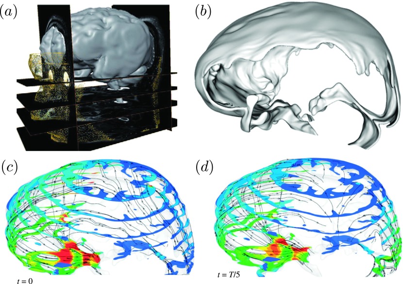
FIG. 10.
Cerebrospinal fluid dynamics in the human cranial subarachnoid space: (a) Anatomic MRI images of the cranial space. (b) Rendering of a reconstructed patient-specific cranial SAS. (c) and (d) Computed CSF velocity magnitude contours at cross sections of the cranial SAS at selected points in time within one cardiac cycle. Reprinted with permission from Gupta et al., “Cerebrospinal fluid dynamics in the human cranial subarachnoid space: An overlooked mediator of cerebral disease. I. Computational model,” J. R. Soc., Interface 7, 1195–1204 (2010). Copyright 2010 Royal Society Publishing.

