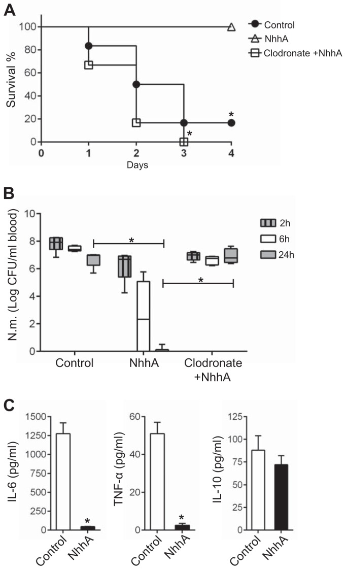
FIG 8 .

NhhA-Mφ prevent bacterial dissemination in vivo. CD46+/+ mice (n = 6 to 8) were treated i.p. with 50 nM heat-inactivated (control) or native NhhA for 3 days prior to being challenged i.p. with FAM20 (108 CFU). To deplete Mo/Mφ, some mice were treated with clodronate-containing liposomes (200 µl i.p.) 4 days prior to NhhA treatment, and depletion was confirmed by monitoring of stained blood smears. (A) Survival of mice at indicated time points postinfection. *, P < 0.05 versus the control group using the Mann-Whitney U test. (B) Bacterial levels in the blood at the indicated postinfection time points. The detection limit is 500 CFU/ml blood. Box plots are divided into upper quartiles and lower quartiles by medians. Error bars indicate the minimum and maximum of data values. *, P < 0.05 using paired Student’s t test. (C) Levels of IL-6, TNF-α, and IL-10 in the serum at 24 h postinfection were determined by ELISA. The data represent the mean ± standard deviation. *, P < 0.05 compared with the control group using paired Student’s t test. The results in panels A to C represent data pooled from three experiments.
