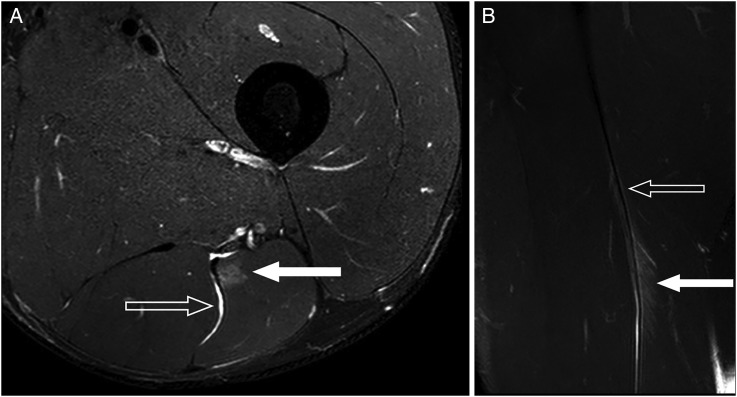
Figure 1.
MRI appearance of injury at muscle-tendon junction of the intramuscular tendon showing characteristic feather-like appearance. (A) Axial fat suppressed image demonstrates intact intramuscular tendon rachis but myofibrillar tearing at the interface with the tendon (solid arrow). Blood products track along the medial margin of biceps coming in contact with the sciatic nerve and separating semitendinosis (open arrow). (B) Coronal fat suppressed images demonstrate classic feather-shaped appearance caused by myofibril disruption with oedema and blood fluid products tracking between the torn muscle fibres (solid arrow). Note that the intramuscular tendon has preserved its linear low signal appearance (open arrow).

