Fig. 3.

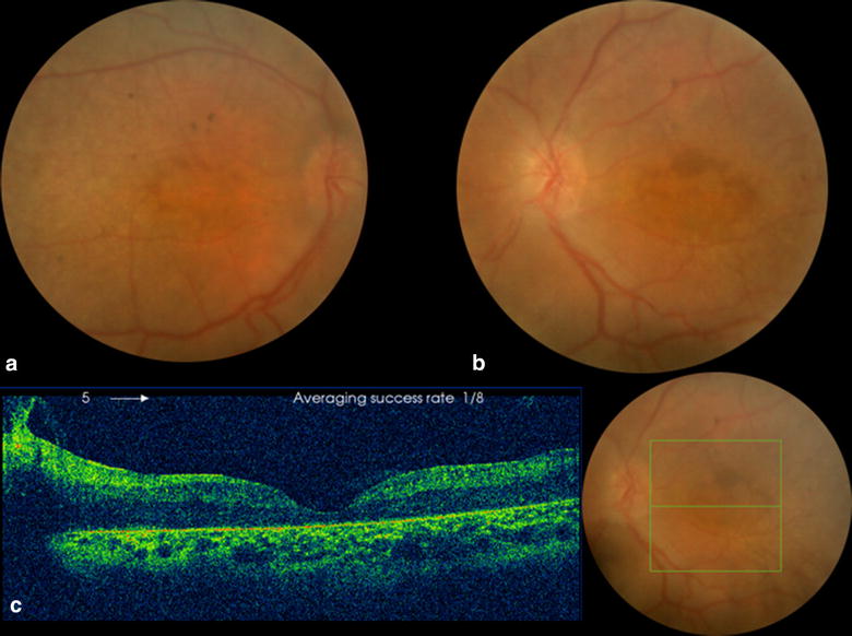
Fundus Photo (OU) and OCT (OS) of the 12 year old male sibling. a, b Fundus photos showing foveal atrophy, nummular pigmentations and moderate vascular attenuation. c OCT showing decreased foveal thickness

Fundus Photo (OU) and OCT (OS) of the 12 year old male sibling. a, b Fundus photos showing foveal atrophy, nummular pigmentations and moderate vascular attenuation. c OCT showing decreased foveal thickness