Fig. 3.

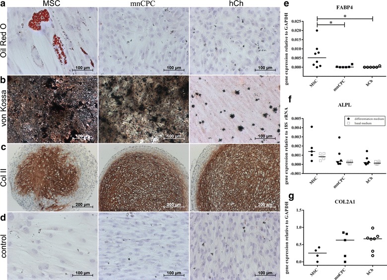
The differentiation potential of mnCPC was compared to hCh and MSC and induced by growth factor supplemented differentiation media over a period of 21 (adipogenic and osteogenic differentiation) and 28 days (chondrogenic differentiation). Only MSC, cultured in adipogenic differentiation medium, exhibited fatty vacuoles positive with Oil red O staining (a). Strong calcium deposition—stained with von Kossa stain—was shown for MSC and mnCPC, only small deposits were found in hCh (b). In the pellet culture for chondrogenic differentiation, collagen type II was detected in all samples (c). All control groups, cultured in basal medium, were negative (d). The respective gene expression analysis (e–g) supported the findings visualised by histological and immunohistochemical staining. Graphs show individual values with median, n = 4–8 donors for each cell type, *p ≤ 0.05. Representative images were chosen
