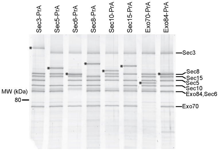
Figure 1.
Purification of intact yeast exocyst complexes. Purified complexes were separated by SDS-PAGE and visualized by Krypton staining (Thermo Scientific). The asterisk corresponds to the PrA-tagged exocyst subunit used as purification handle (shifts the protein molecular weight by 25 kDa). Both the Sec3 and Exo84 protein bands often migrate as multiple species due to phosphorylation, which appear as slightly smeared bands on SDS-PAGE. The resuspension buffer used was 50 mM Hepes pH 7.4, 300 mM NaCl, plus protease inhibitors. Full-size images for this and most gels in Figures 2–5 are shown in Supplementary Data Set 1.

