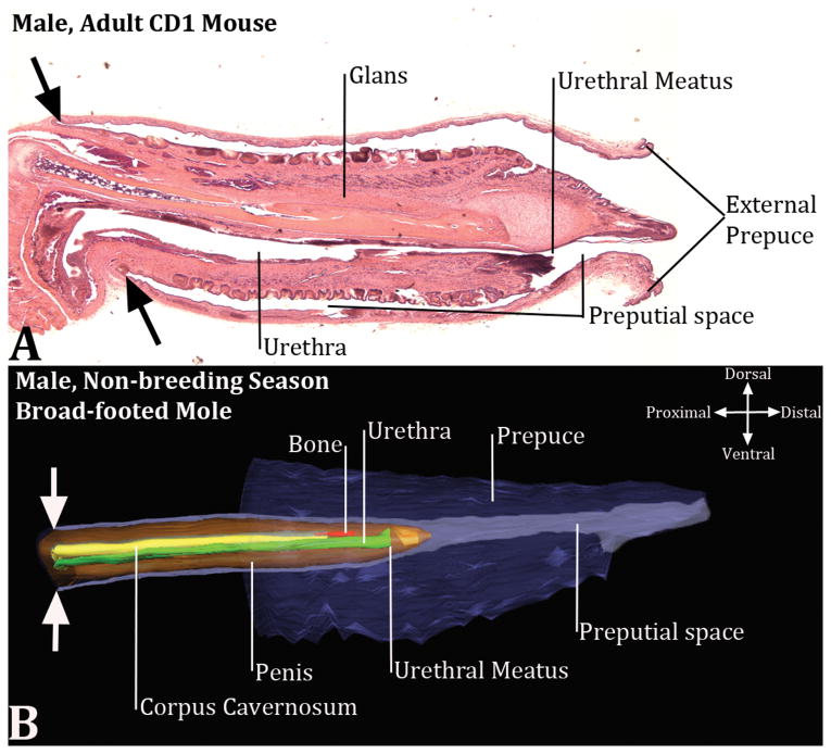
Figure 3.
Mid-sagittal section of the mouse penis (A) and a three dimensional reconstruction of the broad-footed mole external genitalia (B) and associated preputial space. Much of the mouse prepuce has been removed (A), but the fact that the mouse penis is housed within the preputial space is clearly evident. Note the continuity of the inner preputial epithelium with penile surface epithelium denoted by the large black arrows. (B) The three dimensional reconstruction of the broad-footed mole external genitalia, which also indicates that the penis is an “internal organ” housed within the preputial space. The large opposed white arrows in (B) indicate the junction between the external part of the penis that resides within the preputial space and the internal part of the penis that lies deep to the preputial space. In (B) red denotes the os penis, green denotes the urethra, orange denotes the penis, yellow denotes the corpus cavernosum, purple denotes prepuce and preputial space.

