Figure 7.
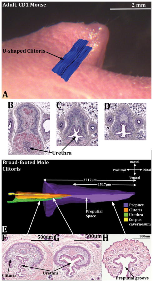
Images demonstrating the “internal” position of the mouse (A) and broad-footed mole clitoris (E) as well as the changing position of the urethra in both species on a proximal/distal basis (B-D and F-H). In (A) a three dimensional reconstruction of the U-shaped clitoral lamina (blue) is superimposed on a side view of adult female mouse external genitalia. Sections B-D in proximal to distal order illustrate the changing position of the urethra relative to the clitoral lamina. Note also that sections (B & C) contain a stand-alone urethra. In (D) a complex epithelium is present in which the clitoral lamina and urethra are fused together. In (E), a three dimensional reconstruction of the broad-footed mole clitoris, the internal position of the clitoris in evident along with its color-coded internal constituents. In sections (F-H), taken at the positions so indicated, the stand-alone urethra is seen (F) as well as the preputial space (G) where the epithelium of the clitoral lamina and urethra are fused. (H) is a section through the female prepuce only showing the preputial groove. CC=corpus cavernosum.

