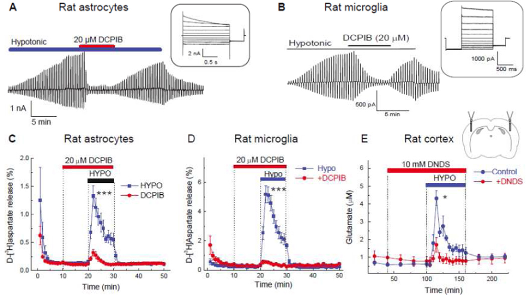
Fig. 2. Representative electrophysiology recordings of VRAC currents and swelling activated glutamate release in brain cells in vitro, and in brain cortex in vivo.
A, Whole-cell recordings of Cl− currents primary rat astrocytes exposed to hypoosmotic medium (−60 mOsm) with symmetrical concentrations of Cl− in bath and pipette solutions. In the main panel, the time course Cl− current development was recorded in response to brief alterative voltage steps to +/−40 mV from the holding potential of 0 mV. In the inset, Cl− currents in response to voltage steps from −100 to +100 mV in 20 mV increments. Note outward rectification and time-dependent current inactivation at high positive potentials. Reproduced with permission from I.F. Abdullaev et al. (2006). B, Whole-cell recordings of VRAC Cl− currents performed in rat primary microglial cells. The conditions and composition of experimental solutions were similar to those used in (A). Reproduced with permission from T.J. Harrigan et al. (2008). C, Swelling activated glutamate release from primary rat astrocytes traced with its non-metabolizable analogue d-[3H]aspartate. Astrocytes were superfused with isoosmotic or hypoosmotic (−100 mOsm) media in the presence or absence of the VRAC blocker DCPIB as indicated. ***p<0.001, effect of DCPIB on glutamate release in swollen cells. Modified with permission from I.F. Abdullaev et al. (2006). D, Swelling activated glutamate release from primary rat microglial cells measured with the non-metabolizable glutamate analogue d-[3H]aspartate. The experimental conditions were identical to those shown in (C). ***p<0.001, effect of DCPIB on glutamate release in swollen cells. Modified with permission from T.J. Harrigan et al. (2008). E, Hypoosmotic stimulation of glutamate release from the rat cortical tissue measured using a microdialysis approach. Cortex was perfused with isoosmotic or hypoosmotic artificial cerebrospinal fluid additionally containing the low affinity VRAC blocker DNDS, which is well tolerated in vivo. Collected microdialysis samples were analyzed off-line for the presence of L-glutamate and other amino acids using HPLC. *p<0.05, effect of DNDS on glutamate release under hypoosmotic conditions. Reproduced from R.E. Haskew-Layton et al. (2008) under the Creative Commons Attribution (CC BY) license.

