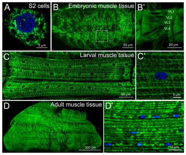
Figure 1. Mitochondrial distribution in Drosophila muscle tissues.
(A–D′) Confocal micrographs of mitochondria immunostained with an antibody against the IMM ATP synthase complex (green) in the indicated cell types. Nuclei are stained with DAPI (blue). (A) An assortment of mitochondrial shapes are observed in S2 cells plated on ConA. (B, B′) Mitochondria are abundant and uniformly distributed in embryonic muscle tissue (B). High magnification of the ventral longitudinal muscles 1–4 (also designated 6, 7, 12, and 13) shows an overall ubiquitous mitochondrial distribution with a slight accumulation at the myofiber ends (B′). (C, C′) In the contractile muscles of third instar larvae, ventral longitudinal muscles 1 and 2 (12 and 13) accumulate around nuclei and adopt an overall striated appearance (C). Tubular mitochondria can be viewed at the surface of the muscle (C′). Mitochondria appear homogeneous in the adult thoracic flight muscle (D) and adopt a round appearance at higher magnification (D′). All images were acquired using a Zeiss 700 confocal microscope and processed using ImageJ and Adobe Photoshop software. Scale bars are indicated.

