
FIG. 4.
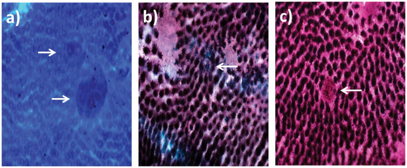
Representative images of (A) methylene-blue stained ACF, (B) sialomucin-expressing ACF, and (C) MDF observed in colons of AαC-fed mice (magnification 40×).
Official websites use .gov
A
.gov website belongs to an official
government organization in the United States.
Secure .gov websites use HTTPS
A lock (
) or https:// means you've safely
connected to the .gov website. Share sensitive
information only on official, secure websites.
Representative images of (A) methylene-blue stained ACF, (B) sialomucin-expressing ACF, and (C) MDF observed in colons of AαC-fed mice (magnification 40×).