Fig. 2.

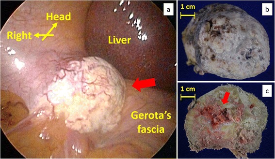
A laparoscopic image of the right upper abdomen and photographs of the excised specimen. a In the right upper retroperitoneum, a white and hard mass was observed next to the right hepatic lobe and the right kidney. b The excised specimen presented calcified mass. c Division surface of the specimen presented mainly calcification and partially red soft tissues
