Fig. 3.

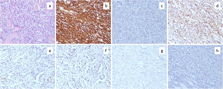
Microscopic findings of the specimen. a An image of hematoxylin-eosin staining. Spindle cells with low cytological atypia densely proliferated in much hyalinizing collagen fiber. b–h Images of immunoreactivity for CD34, Bcl-2, vimentin, SMA, S-100, p53, and Ki-67 are shown, respectively
