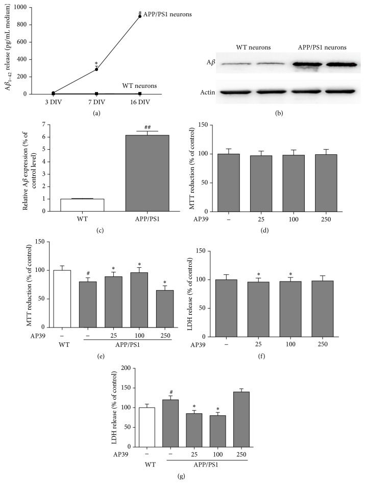
Figure 3.
Cytoprotective effects of AP39 in APP/PS1 neurons. (a) A time course shows that increasing levels of Aβ 42 are released into the culture media of neurons from transgenic mice but not their WT littermates. (b) Aβ was overexpressed in neurons from APP/PS1 mice compared to Aβ expression in neurons from their WT littermates. (c) Representative of Aβ blots is shown with quantification. (d) The effects of AP39 (25–250 nM) treatment for 24 h on cell viability in WT neurons. AP39 alone did not affect MTT conversion. (e) The effects of AP39 (25–250 nM) on MTT conversion in APP/PS1 neurons. There was a decrease in MTT conversion in the APP/PS1 neurons compared to the WT neurons; these effects were attenuated by AP39. (f) After treatment of WT neurons with AP39 for 24 h, AP39 alone did not affect LDH release in the cellular medium. (g) The effects of AP39 on LDH release from APP/PS1 neurons. ∗ P < 0.05, versus the APP/PS1 group; # P < 0.05, the APP/PS1 group versus the WT group.

