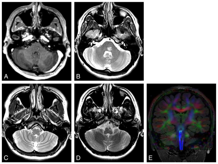
Fig. 1.
Demyelination. Patient presented with horizontal diplopia. Axial contrast T1-weighted image (A) shows a ring enhancing mass in the left paramedian pons. Axial T2-weighted image (B–D) reveals hyperintensity of this demyelinating lesion (B) that decreased over 2 months with steroid treatment (not shown). Image through the inferior olive at presentation (C) shows subtle hyperintensity in the ipsilateral inferior olive consistent with early HOD, with hypertrophy developing 10 months later (D). Tractography superimposed on coronal color FA map (E) shows truncation of the ipsilateral central tegmental tract.

